
Manahil Mubeen, MBBS; Syeda Neha Ahmed, MBBS; Hania Rizwan, MBBS; Karan Bansari, MBBS; Hassan Saleem, MBBS
WMJ. 2026;125(3):340-342. Published August 11, 2026.
Irreversible vision loss resulting from retinal ganglion cell degeneration, such as that occurring in glaucoma, optic neuropathies, and retinitis pigmentosa, continues to present profound clinical challenges. Globally, more than 80 million individuals are affected by glaucoma alone,1 with projections rising to 111.8 million by 2040,2 making retinal nerve damage one of the leading causes of irreversible blindness. Similarly, macular degeneration affects approximately 8.7% of the global population, with the number of affected individuals expected to increase from approximately 196 million in 2020 to 288 million by 2040.3 This growing burden underscores the need for regenerative strategies for retinal neurodegenerative diseases.
Current therapies, such as intraocular pressure-lowering treatments and neuroprotective agents, can slow disease progression but cannot regenerate lost retinal ganglion cells or reverse established neural damage. Mammalian retinas lack significant endogenous regenerative capacity because Müller glia (MG), the retina’s intrinsic support cells with latent progenitor potential, fail to reenter the cell cycle and differentiate into neurons after injury because of epigenetic and signaling constraints.4 In contrast, zebrafish and amphibians mount robust MG-mediated regenerative responses through neurogenic pathways such as Wnt/β-catenin and Ascl1a/Dkk signaling.5-7
Recent advances identify Prospero-related homeobox 1 (Prox1), a homeodomain transcription factor critical for neural development and lymphatic endothelial differentiation, as a novel extracellular barrier to retinal regeneration in mammals.4 In mammalian retinal injury models, Prox1 protein accumulates in MG through intercellular transfer from neighboring retinal interneurons (bipolar and amacrine cells) and suppresses MG proliferation and reprogramming into retinal progenitor cells, thereby arresting the regenerative response.1,4,8 Recent studies have restored MG proliferative capacity and differentiation into retinal neurons, including photoreceptors and retinal ganglion cell-like cells, by blocking Prox1 transfer using an anti-Prox1 nanobody delivered via an adeno-associated virus (AAV) vector. This intervention delayed vision loss in murine retinitis pigmentosa models, highlighting Prox1 as a promising therapeutic target for overcoming intrinsic regenerative barriers in the mammalian retina.1 Below, we examine Prox1 as a potential therapeutic target for retinal regeneration and advocate for increased translational research focused on this novel regenerative strategy.
MULLER GLIA AND RETINOL REGENERATION
MG are radial glial cells that span the entire thickness of the vertebrate retina and provide structural support, neurotransmitter recycling, osmotic balance, and metabolic support to retinal neurons. Retinal regeneration requires MG activation, dedifferentiation, proliferation, and generation of progeny capable of neuronal differentiation and integration into circuits to restore vision.6 Uniquely, MG in cold-blooded vertebrates dedifferentiate in response to injury, downregulate glial markers, upregulate progenitor genes such as Pax6 and Ascl1a, reenter the cell cycle, and generate MG-derived progenitor cells that proliferate and differentiate into lost neuronal subtypes.8 In zebrafish, injury activates Wnt, Notch, Shh, Fgf, HB-EGF, and Yap/Taz pathways to drive progenitor cell proliferation and complete retinal regeneration.7 Avian MG can partially reprogram after excitotoxic damage and persist as undifferentiated progenitors for weeks, unless they are supplemented with exogenous factors.5 By contrast, mammalian MG are unable to spontaneously reprogram after retinal injury without transgenic manipulation or growth factor stimulation. For example, MG in the mouse retina can migrate to sites of injury and upregulate the retinal progenitor cell marker Pax6, but they do not fully reenter the cell cycle or undergo neurogenesis, suggesting incomplete activation of regenerative gene expression programs and the presence of intrinsic inhibitory mechanisms that limit endogenous regeneration.5,8
PROX1 AS A BARRIER TO REGENERATION
Prox1 is a homeodomain transcription factor with well-documented roles in neuronal differentiation and cell fate determination in the central nervous system and retina.4,9-11 In vertebrates, it is essential for lymphatic endothelial identity and the development of specific neuronal lineages, including horizontal interneurons, and plays important regulatory roles in MG differentiation. However, its accumulation in MG after retinal injury acts as a regenerative blockade.9 Notably, Prox 1 undergoes intercellular transfer from neighboring neurons into MG rather than being produced through endogenous transcription, creating a barrier to glia-to-progenitor conversion and representing a rare example of a transcription factor functioning in a non-cell-autonomous manner.4 Within MG, Prox1 suppresses Cyclin D1, E2F5, and PCNA, enforcing G1-phase cell-cycle arrest. Moreover, it blocks activation of proneural factors and represses progenitor markers, including Hbegf and Gadd45a, thereby maintaining MG in a quiescent, gliotic state.4,11
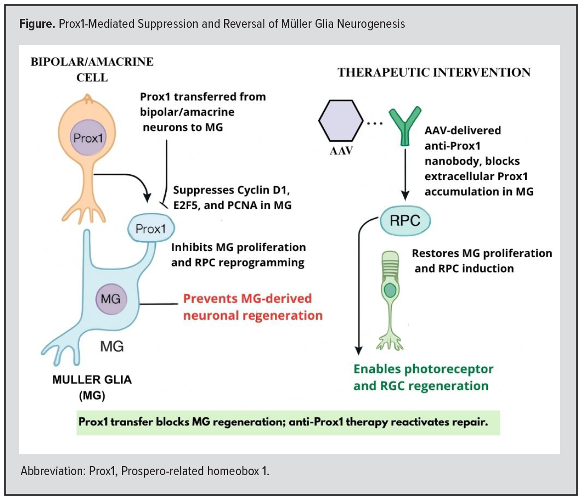 Given this role, genetic disruption or experimental blockade of Prox 1 transfer using AAV-mediated delivery of a secreted anti-Prox1 single-chain antibody reduces MG Prox1 levels, restores MG proliferation, induces expression of retinal progenitor markers (Hes1, Notch1, Gadd45a, Hbegf), and generates nascent photoreceptors and bipolar cells, as demonstrated by EdU incorporation and single-cell RNA sequencing (scRNA-seq).4 In murine models of N-methyl-N-nitrosourea-induced retinal injury and genetic retinitis pigmentosa (eg, rd10 models), anti-Prox1 therapy increased outer nuclear layer thickness, reduced Prox1 protein levels by 60% within 2 weeks, increased MG progenitor cell proliferation and generation of rhodopsin-positive photoreceptors by 45% compared with controls, enhanced scotopic electroretinogram amplitudes by 30 µV, and improved visual acuity by 20% during early disease stages.4,11 Although AAV vector silencing reduced long-term therapeutic efficacy, this approach leverages the resident MG population and may reduce immunogenicity and cellular integration challenges associated with exogenous stem cell therapies while allowing cell type-specific modulation through minimally invasive delivery.12 The Figure illustrates the inhibitory effects of Prox1 transfer on MG regeneration and the restoration of regenerative activity following anti-Prox1 therapy.
TRANSLATIONAL OPPORTUNITIES AND CHALLENGES
Despite strong proof-of-concept data in murine models, translational research remains in its infancy. No human retinal tissue or retinal organoid studies have yet validated Prox1-mediated regeneration. Translating these findings into clinical reality will require careful evaluation of species-specific differences in retinal architecture and biology. Key challenges include validating Prox1-dependent mechanisms in human retinal explants or organoid systems, as sex-based differences, divergent gene expression profiles, and distinct signaling pathways may influence the role of Prox1 in human retinal tissue.
Safety concerns for clinical translation include uncontrolled MG proliferation resulting from overactivation or impaired regulation of MG progenitor cells, potentially leading to disorganized tissue growth, retinal gliosis, ectopic tissue formation, or tumorigenesis.3,5 These risks may be mitigated through tightly controlled, transient gene expression systems11 that limit proliferation once differentiation is achieved. In addition, viral vector delivery can elicit retinal immune responses and inflammation, particularly at high AAV viral loads.10 This challenge may be addressed through the alternative AAV serotypes and engineered capsids designed to enhance tropism and transduction efficiency at lower, safer doses.
Another major concern is aberrant differentiation or off-target effects, in which MG-derived progenitor cells generate inappropriate neuronal subtypes or integrate abnormally into retinal circuits.9 Long-term functional and structural assessments, including electroretinography and optical coherence tomography, can confirm the stability, specificity, and synaptic integration of newly generated neurons. Complementary histological and molecular analyses could further verify neuronal lineage fidelity and circuit connectivity.
Long-term vector expression and specificity also remain unresolved challenges. AAV vector silencing has been shown to reduce the durability of transgene expression, potentially through host-mediated DNA methylation or histone modification, resulting in epigenetic silencing of vector genomes.13 Several alternative delivery strategies may help overcome these limitations. Lentiviral vectors, for example, offer sustained expression through stable genomic integration and have demonstrated prolonged retinal gene expression, although insertional mutagenesis remains a concern.14 Nonviral platforms, including lipid nanoparticles, offer potentially safer and repeatable delivery options and are already being evaluated for retinal applications.15 In addition, next-generation engineered AAV capsids, such as AAV2.7m8 and AAV9-PHP.B, demonstrate improved retinal tropism, reduced interaction with neutralizing antibodies, and enhanced transgene persistence.16 Incorporation of regulatory DNA elements, including ubiquitous chromatin-opening elements and CpG-depleted promoters, may further mitigate epigenetic silencing and prolong vector activity.17 Collectively, improving vector durability, safety, and specificity will be critical to advancing clinical translation. Human retinal organoid models, improved AAV capsid designs, alternative delivery routes such as suprachoroidal administration, and combinatorial regenerative approaches are essential to advance clinical relevance.4
CONCLUSIONS
The growing global burden of irreversible retinal neurodegeneration highlights the urgent need for novel regenerative strategies. The identification of Prox1 as a key inhibitor of MG-driven retinal regeneration provides a promising target for promoting intrinsic retinal repair. Preclinical studies in murine models demonstrate that blockade of extracellular Prox1 can restore MG regenerative capacity, promote neuronal replacement, and preserve visual function. To translate these findings into clinical practice, further research is needed to validate underlying mechanisms in human systems, refine gene delivery platforms, and establish long-term safety. Continued collaborative investigation of Prox1-targeted therapies may ultimately expand treatment options for glaucoma, inherited retinal dystrophies, and retinitis pigmentosa, conditions historically associated with irreversible vision loss – offering hope to millions affected by retinal nerve degeneration..
REFERENCES
- Shan S, Wu J, Cao J, et al. Global incidence and risk factors for glaucoma: a systematic review and meta-analysis of prospective studies. J Glob Health. 2024;14:04252. doi:10.7189/jogh.14.04252
- Tham YC, Li X, Wong TY, Quigley HA, Aung T, Cheng CY. Global prevalence of glaucoma and projections of glaucoma burden through 2040: a systematic review and meta-analysis. Ophthalmology. 2014;121(11):2081-2090. doi:10.1016/j.ophtha.2014.05.013
- Wong WL, Su X, Li X, et al. Global prevalence of age-related macular degeneration and disease burden projection for 2020 and 2040: a systematic review and meta-analysis. Lancet Glob Health. 2014;2(2):e106-e116. doi:10.1016/S2214-109X(13)70145-1
- Lee EJ, Kim M, Park S, et al. Restoration of retinal regenerative potential of Müller glia by disrupting intercellular Prox1 transfer. Nat Commun. 2025;16(1):2928. doi:10.1038/s41467-025-58290-8
- Todd L, Reh TA. Comparative biology of vertebrate retinal regeneration: restoration of vision through cellular reprogramming. Cold Spring Harb Perspect Biol. 2022;14(6):a040816. doi:10.1101/cshperspect.a040816
- Taylor OB, El-Hodiri HM, Palazzo I, Todd L, Fischer AJ. Regulating the formation of Müller glia-derived progenitor cells in the retina. Glia. 2025;73(1):4-24. doi:10.1002/glia.24635
- Lenkowski JR, Raymond PA. Müller glia: Stem cells for generation and regeneration of retinal neurons in teleost fish. Prog Retin Eye Res. 2014;40:94-123. doi:10.1016/j.preteyeres.2013.12.007
- Ramachandran R, Zhao XF, Goldman D. Ascl1a/Dkk/beta-catenin signaling pathway is necessary and glycogen synthase kinase-3beta inhibition is sufficient for zebrafish retina regeneration. Proc Natl Acad Sci U S A. 2011;108(38):15858-15863. doi:10.1073/pnas.1107220108
- Grigoryan EN. Cell Sources for retinal regeneration: implication for data translation in biomedicine of the eye. Cells. 2022;11(23):3755. doi:10.3390/cells11233755
- Hoang T, Wang J, Boyd P, et al. Gene regulatory networks controlling vertebrate retinal regeneration. Science. 2020;370(6519):eabb8598. doi:10.1126/science.abb8598
- Dyer MA, Livesey FJ, Cepko CL, Oliver G. Prox1 function controls progenitor cell proliferation and horizontal cell genesis in the mammalian retina. Nat Genet. 2003;34(1):53-58. doi:10.1038/ng1144
- Pavlou M, Probst M, Kaplan L, et al. AAV-mediated expression of proneural factors stimulates neurogenesis from adult Müller glia in vivo. EMBO Mol Med. 2025;17(4):722-746. doi:10.1038/s44321-025-00209-3
- Das A, Vijayan M, Walton EM, Stafford VG, Fiflis DN, Asokan A. Epigenetic silencing of recombinant adeno-associated virus genomes by NP220 and the HUSH complex. J Virol. 2022;96(4):e0203921. doi:10.1128/JVI.02039-21
- Arsenijevic Y, Berger A, Udry F, Kostic C. Lentiviral vectors for ocular gene therapy. Pharmaceutics. 2022;14(8):1605. doi:10.3390/pharmaceutics14081605
- Chambers CZ, Soo GL, Engel AL, et al. Lipid nanoparticle-mediated delivery of mRNA into the mouse and human retina and other ocular tissues. Transl Vis Sci Technol. 2024;13(7):7. doi:10.1167/tvst.13.7.7
- Dalkara D, Byrne LC, Klimczak RR, et al. In vivo-directed evolution of a new adeno-associated virus for therapeutic outer retinal gene delivery from the vitreous. Sci Transl Med. 2013;5(189):189ra76. doi:10.1126/scitranslmed.3005708
- Zhang F, Frost AR, Blundell MP, Bales O, Antoniou MN, Thrasher AJ. A ubiquitous chromatin opening element (UCOE) confers resistance to DNA methylation-mediated silencing of lentiviral vectors. Mol Ther. 2010;18(9):1640-1649. doi:10.1038/mt.2010.132